___
PRP Therapy for Joint Pain · Ann Arbor, Michigan

Your Body Already Knows How to Heal. We Amplify It.

Dual-centrifuged, ultrasound-guided Platelet-Rich Plasma therapy: the most advanced regenerative protocol available for joint pain, arthritis, and sports injuries. No surgery. No steroids. Your own biology, concentrated and precisely delivered.
  • Dual-Centrifuge PRP
  • Ultrasound-Guided Injections
  • Cash Pay · HSA/FSA · Financing
  • Ann Arbor, Michigan
  • Exosome Upgrade Available
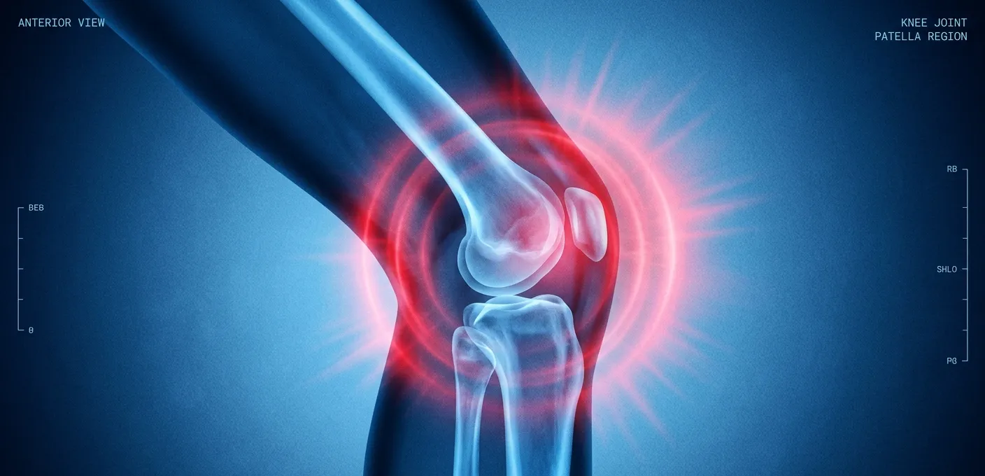
PRP knee joint therapy for pain relief and regeneration
___
What Is PRP Therapy?

Regenerative Medicine, Not Symptom Management.

Platelet-Rich Plasma therapy harnesses the concentrated healing power of your own blood. A small sample is drawn, processed through our dual-centrifuge protocol, producing a platelet concentration 5–7× above baseline, then precisely injected into the affected joint under real-time ultrasound guidance.

Platelets release growth factors, including PDGF, TGF-β, VEGF, and IGF-1, that signal tissue repair, reduce inflammatory cascades, stimulate collagen synthesis, and promote the regeneration of cartilage, tendon, and ligament tissue.

This is not pain management. This is biology-directed repair. For patients seeking an alternative to cortisone injections, viscosupplementation, or surgery, PRP at Arbour Longevity represents the current gold standard in regenerative joint care.

◆ Protocol Standard

Dual-Centrifuge PRP · 5–7× Platelet Concentration · Ultrasound-Guided Delivery

◆ Joints Treated

Knee · Shoulder · Hip · Elbow · Ankle · Foot · Wrist · Hand

◆ Investment

Single Session: $800–$1,200

Series of 3: $2,500–$3,500

Exosome Upgrade: $2,500–$3,500

◆ Payment

Cash Pay · HSA/FSA Accepted · Financing Available

The Evidence

Science-Backed. Clinician-Administered.

PRP therapy is among the most studied regenerative interventions in orthopedic medicine. At Arbour Longevity, we follow evidence-based protocols, using dual-centrifuge processing to maximize platelet yield and ultrasound guidance to ensure precise intra-articular delivery every time.

5–7×

Platelet Concentration

Dual-centrifuge processing produces 5–7× baseline platelet concentration, the clinically effective threshold for tissue regeneration.

73%

Patients Report Improvement

In peer-reviewed knee OA studies, over 73% of PRP patients reported significant pain and function improvement at 12-month follow-up.

100%

Ultrasound-Guided

Every Arbour Longevity PRP injection is performed under real-time ultrasound guidance, ensuring precise intra-articular placement, not approximation.

6–12

Months Duration

Clinical improvements from PRP typically persist 6–12+ months, with many patients experiencing durable results from a single series.

The Arbour Longevity PRP Protocol

Dual-Centrifuge. Ultrasound-Guided. Precision From Start to Finish.
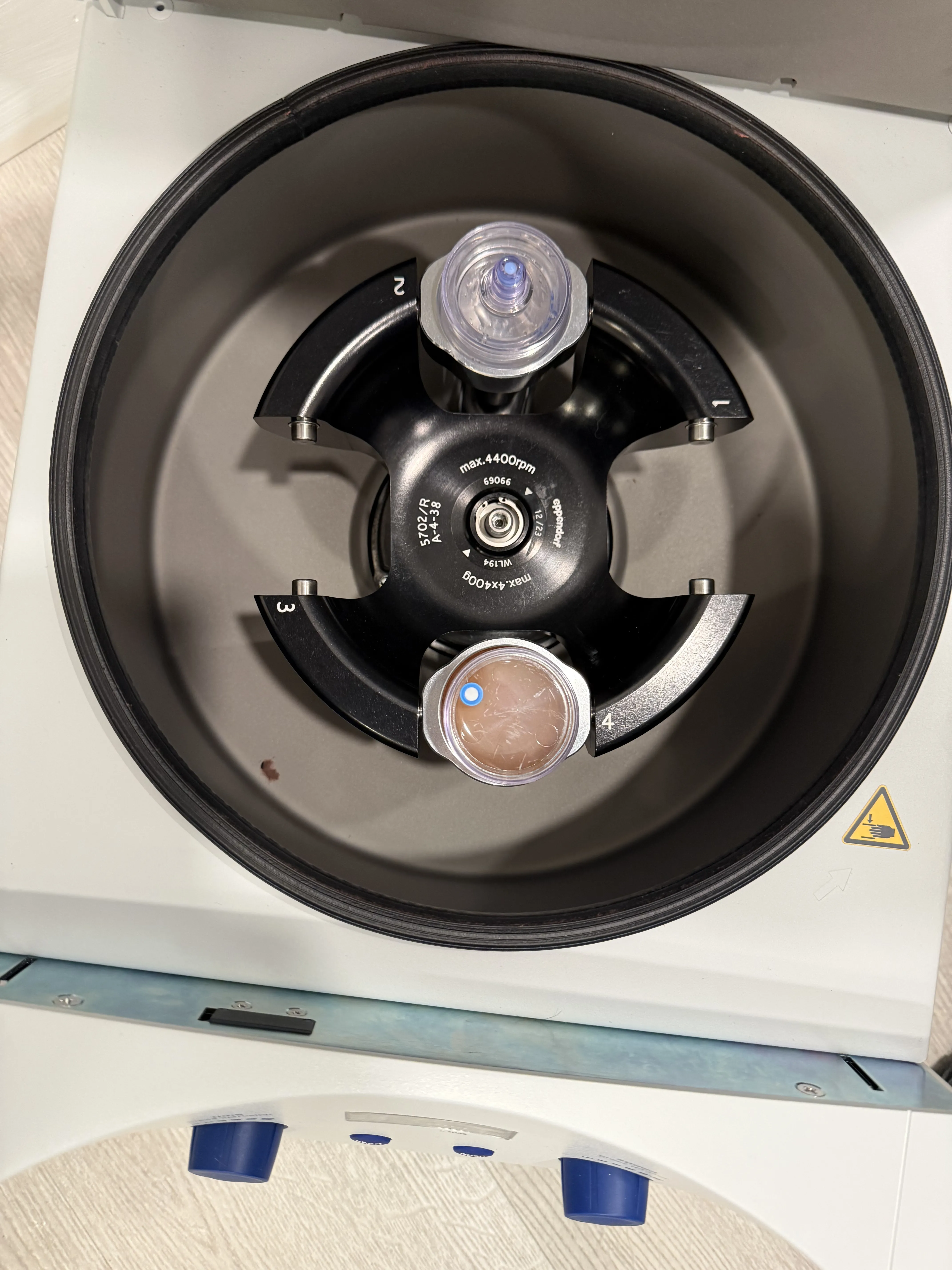
Professional treatment at Arbour Longevity Ann Arbor
01

Consultation

Full clinical assessment: imaging review, functional evaluation, candidacy determination. In-person at our Ann Arbor clinic.
02

Blood Draw

30–60 ml blood drawn in-office, sterile collection into specialized PRP tubes with anticoagulant.
03

Dual Centrifuge

Two-stage centrifugation separates and concentrates platelets to 5–7× baseline. Superior to single-spin protocols.
04

US-Guided Injection

Real-time ultrasound confirms needle placement within the joint space. Precise intra-articular delivery every time.
05

Recovery & Follow-Up

Brief activity modification 48–72 hrs. Follow-up at 4–6 weeks. A series of 3 is recommended for optimal outcomes.

Joints & Conditions Treated

One Protocol. Every Joint. Precisely Delivered.
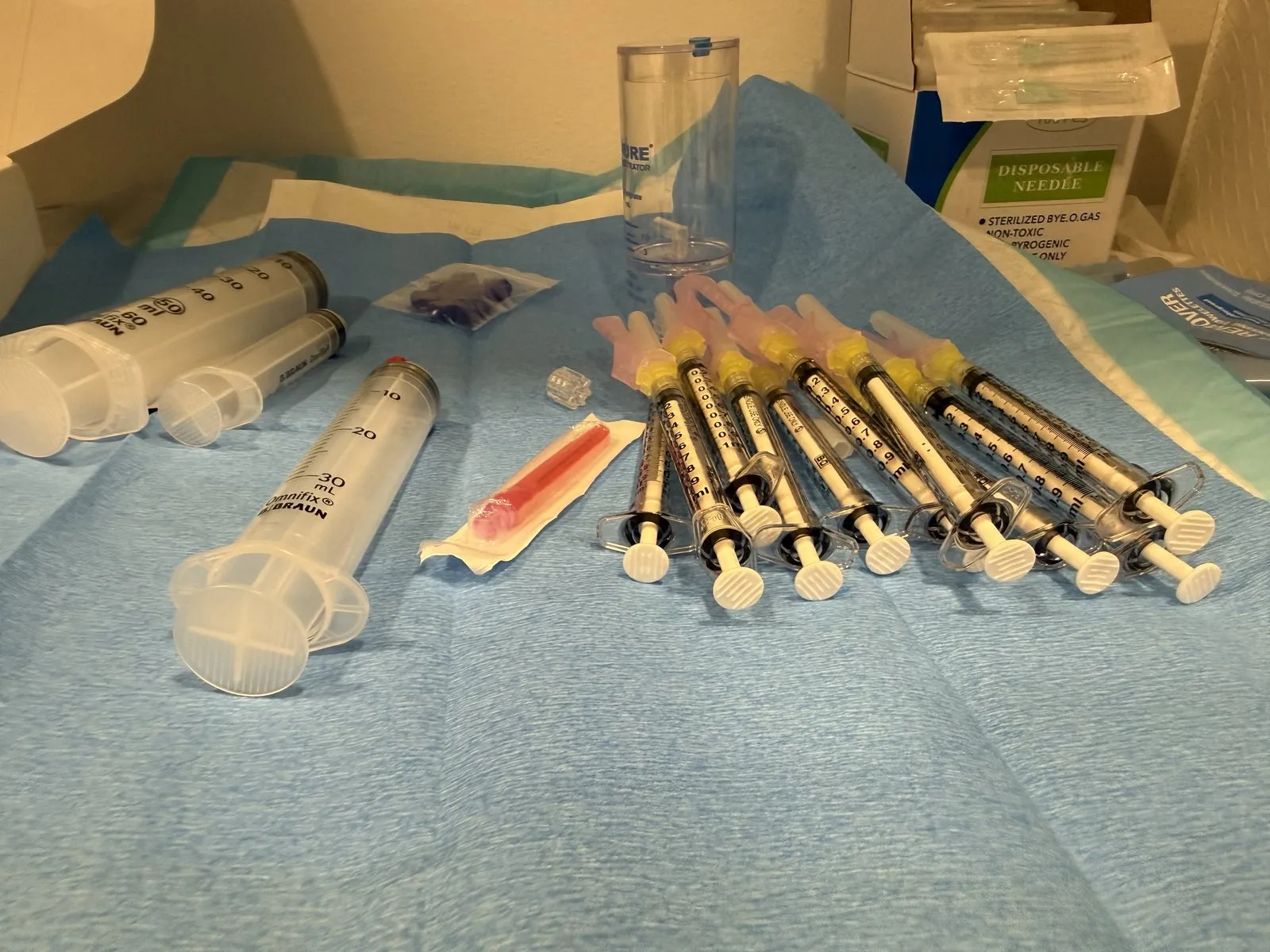
PRP preparation for joint pain therapy

Highest SEO Priority

Knee Joint

Osteoarthritis, meniscal degeneration, ACL/PCL support, patellar tendinopathy, and post-surgical recovery. Arbour Longevity's highest-demand PRP indication.

Common Sports Injury

Shoulder

Rotator cuff tears and tendinopathy, labral degeneration, subacromial impingement, AC joint arthritis, and biceps tendon pathology.

Weight-Bearing Joint

Hip

Hip osteoarthritis, labral tears, greater trochanteric bursitis, piriformis syndrome, and hip flexor tendinopathy.

Overuse Injury

Elbow

Lateral epicondylitis (tennis elbow), medial epicondylitis (golfer's elbow), and common extensor tendon pathology.

Extremity Joints

Ankle, Foot & Wrist

Ankle OA, Achilles tendinopathy, plantar fasciitis, wrist arthritis, De Quervain's tenosynovitis, and finger/thumb joint pain.

Premium Add-On

HA Knee Filler

Hyaluronic acid viscosupplementation for knee joint lubrication: a powerful complement to PRP or standalone treatment for early-stage OA.

Patient Candidacy

Are You a Candidate for PRP Therapy?

PRP is highly effective for the right patient, and our candidacy assessment ensures every protocol is clinically appropriate. Here is what our clinician evaluates before recommending treatment.

Ideal Candidates: PRP Works Best When

Joint pain lasting 3+ months that hasn't resolved with conservative care
Mild to moderate osteoarthritis (Kellgren-Lawrence Grade 1–3)
Tendon or ligament injuries: partial tears, tendinopathy, chronic sprains
Athletes and active patients seeking faster, non-surgical recovery
Patients seeking to delay or avoid joint replacement surgery
Patients who have plateaued on cortisone or NSAID therapy
Generally healthy individuals with adequate platelet function

Relative Contraindications: Requires Evaluation

Active infection at or near the injection site
Severe osteoarthritis (Kellgren-Lawrence Grade 4): exosome therapy may be preferred
Active blood-thinning medication (requires evaluation)
Platelet disorders or active hematologic conditions
Systemic corticosteroid use within the past 2 weeks
Active cancer or immunosuppressive therapy

Not sure if you qualify? Our candidacy assessment takes 3 minutes and gives you a personalized recommendation before you book a consultation.

Take Candidacy Assessment →
___
Premium Upgrade

Beyond PRP. Exosome Therapy.

For patients with advanced joint degeneration, failed prior PRP, or who want the most powerful regenerative intervention available, exosome therapy delivers a concentrated payload of over 200 growth factors, cytokines, and regenerative signaling molecules.

It is the next frontier of joint regeneration, and it is available at Arbour Longevity.

$2,500–$3,500

Per Session · Can Be Combined With PRP

200+ growth factors vs. the ~30 in PRP
No blood draw required: off-the-shelf administration
Ideal for KL Grade 3–4 osteoarthritis
Can be combined with PRP for maximum regenerative effect
Also available for: hair, aesthetics, and sexual wellness
Take Candidacy Assessment →

PRP vs. Exosome: Which Is Right for You?

The right choice depends on the severity of your condition, your goals, and your budget. We guide every patient to the protocol that gives them the best outcome for their specific situation.

Our clinician reviews your imaging, symptom history, and prior treatments before making any recommendations. Exosome therapy is never sold as a default; it is recommended when the evidence supports it.

Start Candidacy Assessment

Investment & Pricing

Transparent Pricing. No Hidden Costs.

All PRP services at Arbour Longevity are cash-pay. HSA and FSA funds are accepted. Financing is available through our patient financing partner for qualified applicants.

Single Session

PRP Injection

$800–$1,200

Per Joint · Per Session

Dual-centrifuge protocol
Ultrasound-guided delivery
Post-procedure care plan
Platelet disorders or active hematologic conditions
4–6 week follow-up included
Schedule Single Session

Most Recommended

Series of 3

$2,500–$3,500

Complete Series · Most Recommended

3 dual-centrifuge sessions
Ultrasound-guided every session
Spaced 4–6 weeks apart
Full progress monitoring
Save vs. single session pricing
Schedule Series

Premium Upgrade

Exosome Therapy

$2,500–$3,500

Per Session · Advanced Regeneration

200+ regenerative growth factors
Ultrasound-guided delivery
Ideal for Grade 3–4 OA
Can be combined with PRP
No blood draw required
Learn About Exosomes

Financing Available: Monthly payment plans available through our patient financing partner. Most patients qualify. Ask about financing during your consultation.  |  HSA / FSA: Accepted for all PRP and regenerative services.

Frequently Asked Questions

What Patients Ask Us Most.

What makes dual-centrifuge PRP different from standard PRP?
+
-

Most PRP clinics use a single-spin centrifuge, producing 2–3× platelet concentration, the minimum therapeutic threshold. Arbour Longevity's dual-centrifuge protocol produces 5–7× baseline concentration, delivering a significantly higher density of regenerative growth factors to the treatment site. The difference in platelet yield directly correlates with clinical outcomes.

Why is ultrasound guidance important for PRP injections?
+
-

Studies show that blind (palpation-guided) joint injections miss the intra-articular space in 20–40% of cases, depending on the joint. When PRP is injected outside the joint, its therapeutic effect is dramatically reduced. Every Arbour Longevity injection is performed under real-time ultrasound visualization, ensuring the PRP is delivered exactly where it needs to be, every time.

How many PRP sessions will I need?
+
-

We recommend a series of 3 injections spaced 4–6 weeks apart for most patients; this is the protocol supported by the strongest clinical evidence. Some patients experience meaningful improvement after a single injection, while others with more advanced degeneration may benefit from a second series at 6–12 months.

Is PRP covered by insurance?
+
-

PRP is not covered by most insurance plans, as it is classified as investigational by many carriers. Arbour Longevity operates as a cash-pay practice for regenerative services. We accept HSA and FSA funds, and patient financing is available for qualified applicants. Many patients find the cost of a PRP series compares favorably to the cumulative cost of ongoing cortisone injections, physical therapy, and eventual surgical consultation.

How does PRP compare to cortisone injections?
+
-

Cortisone provides rapid anti-inflammatory relief, typically lasting 6–12 weeks, but does not address the underlying tissue damage and can accelerate cartilage degradation with repeated use. PRP works differently: it initiates a regenerative response that builds over 4–12 weeks and may produce durable improvements lasting 6–18 months or longer. PRP treats the cause. Cortisone masks the symptom.

What is the recovery after a PRP injection?
+
-

Most patients experience mild soreness and swelling at the injection site for 24–72 hours, this is the normal inflammatory phase of the healing response. Activity modification is recommended for 48–72 hours. Most patients return to normal activities within a few days, with gradual return to sport or strenuous activity guided by symptom response over 2–4 weeks.

PRP Therapy · Ann Arbor

Your Joints Can Heal. Let's Find Out If PRP Is Right for You.

Schedule a regenerative medicine consultation at Arbour Longevity: we'll review your imaging, assess your candidacy, and recommend the protocol that gives you the best outcome for your specific condition.

  • Cash Pay
  • HSA / FSA Accepted
  • Financing Available
  • Ann Arbor, MI
Schedule Regenerative Consultation

Clinician-led · Accepting new patients

Conditions We Treat With PRP

Platelet-rich plasma is used across a range of joint and tendon conditions, but the strength of the supporting evidence is not the same for all of them. The summaries below describe what each condition is, how PRP is thought to act on that particular tissue, what published research actually shows, and where it tends to fall short. An important disclosure first: PRP for orthopedic and musculoskeletal conditions is not FDA-approved and is considered investigational for most of these indications. Blood-derived products of this kind are regulated as minimally manipulated autologous tissue rather than as an approved drug or device, response varies considerably between individuals, and treatment is generally not covered by insurance. Nothing here is a substitute for an individual evaluation.

Knee Osteoarthritis

Knee osteoarthritis is the gradual breakdown of articular cartilage alongside low-grade synovial inflammation, subchondral bone remodeling, and a measurable decline in the quality of joint fluid. Cartilage has no direct blood supply, so once it thins the joint has limited capacity to repair itself. Prior meniscal injury, malalignment, excess load, and inflammatory signaling all accelerate the process.

PRP is thought to act on the joint environment more than on cartilage itself. Concentrated platelets release growth factors including PDGF, TGF-beta, VEGF and IGF-1, which laboratory and early clinical work suggests may prompt synovial cells to produce more hyaluronic acid, modulate inflammatory cytokines such as IL-1beta, and improve the lubricating quality of synovial fluid. The realistic aim is a quieter, better-cushioned joint, not regrown cartilage.

Knee OA is by a wide margin the most studied PRP indication. Multiple randomized controlled trials and several meta-analyses have reported greater improvement in pain and function scores at six to twelve months compared with hyaluronic acid and with saline injection, most consistently in mild-to-moderate radiographic disease. That said, preparation methods differ between studies and at least one large, well-designed trial found no advantage over saline, so professional societies still describe the evidence as encouraging rather than settled.

A typical protocol is two to three injections spaced roughly two to four weeks apart, with formal reassessment at about three months. Many patients report a soreness flare for several days, gradual change over four to eight weeks, and peak benefit near the three-month mark. When patients do respond, benefit commonly lasts six to twelve months, and some choose to repeat treatment on roughly an annual basis.

Honest assessment: Response is generally weaker in bone-on-bone (Kellgren-Lawrence grade 4) arthritis, significant limb malalignment, large recurring effusions, or inflammatory arthritis. PRP is not a substitute for joint replacement in end-stage disease, and some patients notice no change at all.

Rotator Cuff & Shoulder Tendinopathy

The rotator cuff is a group of four tendons that centers and stabilizes the shoulder. What is casually called tendinitis is usually tendinosis: repeated loading outpaces repair, collagen fibers become disorganized, and the tissue near the tendon's insertion, where blood supply is naturally sparse, settles into a stalled healing state rather than an actively inflamed one. Partial-thickness tearing often develops within that degenerative tissue.

In tendon, PRP is thought to work by delivering a concentrated growth-factor signal directly into tissue that has stopped responding. Evidence suggests this may recruit tenocytes, support new collagen synthesis, and encourage small-vessel ingrowth. Placement matters: the injection is generally targeted into or immediately around the degenerative tendon rather than into the subacromial bursa, because bursal placement does not deliver the signal to the diseased tissue.

The published evidence here is moderate rather than strong. Randomized trials and pooled analyses support PRP for chronic cuff tendinopathy and partial-thickness tears, with several studies showing corticosteroid outperforming PRP in the first weeks but PRP producing better pain and function scores at six to twelve months. Results for PRP as an adjunct to arthroscopic repair of full-thickness tears are genuinely mixed, with some trials reporting lower re-tear rates and others showing none.

Protocols commonly involve one to two injections spaced four to six weeks apart, usually with ultrasound guidance for accuracy. Relative rest for one to two weeks is typically followed by a structured progressive loading program, since tendon outcomes in published studies are consistently better when injection is paired with rehabilitation. Most patients who respond describe change between six and twelve weeks, not days.

Honest assessment: Full-thickness retracted tears, tears with significant muscle atrophy or fatty infiltration, and adhesive capsulitis are unlikely to respond meaningfully and usually require surgical or different management. Patients unable to commit to rehabilitation tend to do poorly regardless of injection quality.

Hip Osteoarthritis & Gluteal Tendinopathy

The hip is a deep, tightly enclosed ball-and-socket joint. Osteoarthritis there follows the same pattern as the knee, with cartilage thinning, capsular tightening, and altered joint fluid, but the anatomy makes it a harder target. Gluteal tendinopathy, often mislabeled trochanteric bursitis, is a separate and very common problem: degeneration of the gluteus medius and minimus tendons at the outer hip, producing lateral pain that worsens with side-lying and stair climbing.

For hip osteoarthritis, PRP is thought to act on synovial inflammation and fluid quality much as it does in the knee. For gluteal tendinopathy, the proposed mechanism is the tendon one, delivering growth factors into degenerative tissue to support a repair response that has stalled. These are different targets and should not be assumed to behave the same way.

Accuracy is the practical issue. Landmark-guided hip joint injections miss the capsule at meaningful rates in published accuracy studies, so intra-articular hip treatment is generally performed under ultrasound or fluoroscopic guidance. The evidence base is also thinner than for the knee: hip OA trials are fewer, smaller, and less consistent, with some showing PRP comparable or modestly superior to hyaluronic acid and others showing no clear separation. Gluteal tendinopathy has somewhat more encouraging randomized data, including trials suggesting better twelve-week and two-year outcomes than corticosteroid injection.

Typical protocols run one to three sessions spaced two to four weeks apart, with reassessment near three months and hip abductor strengthening as a core part of care for tendinopathy. Expect gradual change over six to twelve weeks rather than immediate relief.

Honest assessment: Advanced hip OA with severe joint space loss, hip dysplasia, avascular necrosis, or referred pain originating from the lumbar spine responds poorly. Because hip evidence is weaker than knee evidence, expectations should be set accordingly, and patients already near hip replacement are usually better served by surgical consultation.

Tennis & Golfer's Elbow (Lateral and Medial Epicondylitis)

Lateral epicondylitis (tennis elbow) and medial epicondylitis (golfer's elbow) involve degeneration where the wrist extensor or flexor tendons attach at the elbow. Despite the -itis suffix, biopsy studies consistently show angiofibroblastic degeneration rather than true inflammation: disorganized collagen, immature blood vessels, and an absence of typical inflammatory cells. That is precisely why anti-inflammatory strategies often fail to produce lasting change in chronic cases.

PRP is thought to be well suited to this tissue because the underlying problem is a failed repair response rather than inflammation. Injecting concentrated platelets into the degenerative enthesis, usually with a peppering or fenestration technique that also creates controlled micro-trauma, is believed to reintroduce the growth-factor signaling needed to restart remodeling.

This is one of the better-supported non-knee indications. Multiple randomized trials and systematic reviews report that PRP outperforms corticosteroid injection at six-month and twelve-month follow-up for chronic lateral epicondylitis, even though steroid provides faster short-term relief. Some placebo-controlled trials show smaller differences than the steroid comparisons, so the effect size is debated, but the direction of the evidence is comparatively consistent. Chronic cases that have already failed bracing, activity modification, and physical therapy appear to be the group that benefits most.

Most protocols use a single injection, with a second considered at six to eight weeks if progress has plateaued. Patients are typically asked to avoid gripping-heavy activity briefly, then begin eccentric wrist strengthening. Soreness for three to seven days is common, and meaningful improvement usually emerges between four and twelve weeks, sometimes continuing to improve out to six months.

Honest assessment: Symptoms driven by cervical radiculopathy, ulnar or radial nerve entrapment, or a complete tendon rupture will not improve with PRP, because the diagnosis is different. Very short-duration symptoms often resolve with conservative care alone and may not justify injection.

Achilles Tendinopathy & Plantar Fasciitis

The Achilles tendon and plantar fascia both carry very high repetitive load with relatively limited blood supply. Achilles tendinopathy occurs either at the mid-portion, roughly two to six centimeters above the heel, or at the insertion, and each behaves differently. Plantar fasciitis, more accurately plantar fasciopathy, is degeneration of the fascia at its heel attachment, producing the characteristic first-step morning pain.

The proposed mechanism is again tendon and fascia repair signaling: growth factors delivered into degenerative tissue to support collagen remodeling where the native healing response has stalled. Ultrasound guidance is commonly used to place the injection into the abnormal tissue rather than around it.

The evidence deserves a candid summary because it splits. Plantar fasciitis has reasonable support, with several randomized trials and meta-analyses reporting better pain scores at three to six months compared with corticosteroid injection, along with the practical advantage of not carrying steroid's fascial rupture and fat pad atrophy risks. Mid-portion Achilles tendinopathy is different: the randomized evidence is genuinely conflicting, with well-conducted placebo-controlled trials showing no significant advantage over saline when both groups performed eccentric exercise, while other studies and registries report benefit. Anyone claiming the Achilles question is resolved is overstating the literature.

Protocols typically involve one to two injections spaced four to six weeks apart, always paired with a progressive loading program, which itself has stronger evidence than any injection for Achilles disease. Post-injection soreness of several days is normal, with response, when it occurs, developing over six to twelve weeks.

Honest assessment: PRP is not appropriate for a complete or high-grade partial Achilles rupture, and injection into that setting can be harmful. Insertional Achilles disease with Haglund deformity, systemic inflammatory arthropathy, and heel pain from nerve entrapment or calcaneal stress fracture respond poorly. Given the conflicting Achilles data, load management should be optimized first.

Sacroiliac Joint & Lumbar Facet Joint Pain

The sacroiliac joints transmit load between spine and pelvis, and the lumbar facet joints are small paired synovial joints guiding motion at each spinal level. Both are true synovial joints with cartilage, a capsule, and dense nerve supply, and both degenerate with age, prior injury, altered mechanics, or after lumbar fusion changes load distribution. Together they account for a meaningful share of chronic low back pain.

The rationale for PRP here mirrors other joints: platelet-derived growth factors are thought to modulate the local inflammatory environment and support the capsular and ligamentous tissue surrounding the joint. Because these joints are small, deep, and irregular, image guidance with fluoroscopy or ultrasound is considered necessary for accurate placement, and diagnostic anesthetic blocks are often used first to confirm the joint is actually the pain generator.

This is the most investigational of the six indications discussed here, and the evidence should be described that way. Published work consists largely of small randomized trials, prospective case series, and short follow-up periods. Some comparative studies of sacroiliac joint PRP versus corticosteroid report longer-lasting relief with PRP, and early lumbar facet and intradiscal work is described as promising, but sample sizes are small, control conditions vary, and independent replication is limited. It would be inaccurate to present spinal PRP as established care.

When it is offered, protocols generally involve one to two guided injections spaced four to six weeks apart, combined with core and hip stabilization rehabilitation. Response, if it comes, typically develops over four to twelve weeks. Reassessment should be explicit, with a clear plan to stop if there is no meaningful change.

Honest assessment: Patients with radicular pain from disc herniation or stenosis, spondyloarthropathy, spinal instability, fracture, or unexplained progressive neurological symptoms need a different workup entirely. Because the evidence here is the thinnest of any indication on this page, spinal PRP is best considered only after standard conservative options and clear diagnostic confirmation.

Candidacy for PRP is assessed individually rather than by diagnosis alone. That assessment usually includes a physical examination, a review of prior treatments, and in many cases a review of existing imaging or new imaging to confirm the pain generator and rule out conditions that would not respond. PRP is generally not appropriate for patients with an active local or systemic infection, active cancer, certain blood disorders including significant thrombocytopenia or platelet dysfunction, or anemia below safe thresholds, and it is typically deferred during pregnancy or when anticoagulation cannot be safely managed. Non-steroidal anti-inflammatory medications are usually held for a period before and after treatment, because they blunt the same inflammatory signaling cascade PRP is intended to activate, and recent corticosteroid injection at the same site may also warrant a delay. Where the evidence is weaker, that will be said plainly during consultation so expectations are set honestly before treatment begins.